ABE-IPSABE HOLDINGABE BOOKS
English Polski
On-line access

Bookstore

0.00 PLN
Bookshelf (0) 
Your bookshelf is empty
An Atlas of Comparative Vertebrate Histology

An Atlas of Comparative Vertebrate Histology

Authors
Publisher Academic Press
Year 01/06/2018
Edition First
Pages 634
Version other
Language English
ISBN 9780124095977
Categories Medicine: general issues
lifetime license
Product available online
Delivery: access code sent by e-mail
E-Mail
order with obligation to pay
Add to bookshelf

Book description

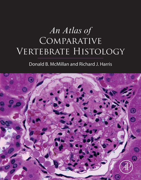
Atlas of Comparative Vertebrate Histology looks at the histology of a wide range of vertebrates, representative of all the major classes and families, with examples ranging from amphioxus to primates. The authors focus their microscope on commonly seen vertebrates as well as 'non-standard' species, such as lamprey, hagfish, dogfish, skate, rock bass, cod, river catfish, toad, amphiuma, leopard and bull frog, garter and brown snake, Coturnix quail and cowbird. The study of comparative histology in the vertebrates helps students and researchers alike understand how various groups have addressed similar problems, opening doors to interesting research possibilities. Not all vertebrates follow the mammalian model of tissue and organ structure. When dealing with unique species, we see some structures taken beyond their 'normal' function. Comparative histology allows us to understand the structural responses underlying the physiology unique to each vertebrate group. Presents the histology of a wide range of vertebrates, representative of all the major classes and families, with examples ranging from amphioxus to primates Includes an image gallery with over 500 flat images and 50+ virtual microscopy slides Contains electronic content features cross linking between text, tables and the image gallery

An Atlas of Comparative Vertebrate Histology

Table of contents


  • An Atlas of Comparative Vertebrate Histology

  • Copyright Page

  • Dedication

  • Contents

  • Introduction

  • Preparing and Studying Tissues for Histology

  • How Big Is Small?

  • Preparing Tissue for Microscopic Examination

  • Fixation

  • Dehydration

  • Embedding

  • Sectioning

  • Mounting

  • Staining and Coverslipping

  • Interpreting Microscopic Images

  • How Microscopes Work?

  • Lens and Image Defects

  • Resolution

  • Electron Microscopy

  • Resolution in Transmission Electron Microscope

  • Notes About Magnifications and Repetition of Images Used in This Book

  • Acknowledgments

  • I. Cells

  • A. The Animal Cell

  • Introductory Study

  • ?Typical? Animal Cell

  • Study of Cellular Structure With the Light Microscope

  • Cytoplasmic Organelles

  • Cytoplasmic Inclusions

  • Nucleus

  • Study of Cellular Structure With the Electron Microscope

  • Cell Membrane (Plasmalemma)

  • Endoplasmic Reticulum and Ribosomes

  • Mitochondria

  • Golgi Complex

  • Lysosomes

  • Centrosomes and Fibrils

  • Cytoplasmic Inclusions

  • Nucleus

  • Freeze Fracture

  • B. Cell Division

  • Mitosis

  • Interphase

  • Prophase

  • Prometaphase

  • Metaphase

  • Anaphase

  • Telophase and Cytokinesis

  • Colchicine, Mitosis, and Karyotypes

  • Duration of the Mitotic Cycle

  • Amitosis

  • Meiosis

  • II. Tissues

  • C. Epithelial Tissues

  • Classification of Epithelia

  • Shape of Epithelial Cells

We also recommend books

Strony www Białystok Warszawa
801 777 223