$23.62 (with VAT)
105.00 PLN / €22.51 / £19.54
Delivery to United States
check shipping prices
Product to order
Delivery 3-4 weeks
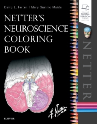
Reinforce your knowledge of neuroanatomy, neuroscience, and common pathologies of the nervous system with this active and engaging learn and review tool! Netter's Neuroscience Coloring Book by Drs. David L. Felten and Mary Summo Maida, challenges you to a better understanding of the brain, spinal cord, and peripheral nervous system using visual and tactile learning. It's a fun and interactive way to trace pathways and tracts, as well as reinforce spatial, functional, and clinical concepts in this fascinating field. More than "just" a coloring book, this unique learning tool offers:
- More than 100 key topics in neuroscience and neuroanatomy, using bold, clear drawings based on classic Netter art.
- Clinical Notes that bridge basic science with health care and medicine.
- Workbook review questions, and bulleted lists throughout to reinforce comprehension and retention.
- Expert Consult(TM) eBook version included with purchase. This enhanced eBook experience allows you to search all of the text, figures, and references from the book on a variety of devices.
More than "just? a coloring book, this unique learning tool offers:
- More than 145 key topics in neuroscience and neuroanatomy, using bold, clear drawings based on classic Netter art.
- Coloring exercises for visual and tactile learning as you trace pathways and tracts, reinforcing spatial, functional, and clinical concepts in this fascinating field.
- A clear organization with 4 major sections: (1) Overview of the nervous system; (2) regional neuroscience; (3) systemic neuroscience; and (4) global neuroscience.
- Three major components for each topic and accompanying illustrations: What is it and what does it do?; Color the most important structures; and What is the functional and clinical significance?
- Text revision based on extensive student feedback.
- New coloring exercises on Endogenous Opioid Systems, Insular Cortex, Prefrontal Cortex, Dementias, Alzheimer's Disease, Posttraumatic Stress, Traumatic Brain Injury (TBI), and Brain Substrates of Addictive Disorders.
- Clinical Notes that bridge basic science with health care and medicine.
- Expanded workbook review questions and bulleted lists throughout to reinforce comprehension and retention.
Enhanced eBook version included with purchase. Your enhanced eBook includes
completed coloring and workbook pages for reference and allows you to access all of the text and figures from the book on a variety of devices.
Netter's Neuroscience Coloring Book
Section 1 Overview of the Nervous System
Chapter 1, Neurons and Their Properties
Neuronal Structure
Types of Synapses
Neuronal Cell Types
Glial Cell Types
Astrocyte Biology
Microglial Biology
Oligodendroglial Biology
The Blood-Brain Barrier
Axonal Transport in the CNS and PNS
Myelination of CNS and PNS Axons
Neuronal Resting Potential
Graded Potentials in Neurons
Action Potentials
Conduction Velocity
Neurotransmitter Release
Multiple Neurotransmitter Synthesis, Release, and Signaling from Individual Neurons
Chemical Neurotransmission
Chapter 2, Brain, Skull and Meninges
Meninges and Their Relationships to the Brain and Skull
Surface Anatomy of the Forebrain: Lateral View
Cerebral Cortex Anatomy and Functional Regions: Lateral View
Cortical Architecture: Brodmann's Areas
Midsagittal Surface of the Brain
Basal Surface of the Brain
Axial and Mid-Sagittal Views of the Central Nervous System
Horizontal (Axial) Brain Sections Showing the Basal Ganglia
Major Limbic Forebrain Structures
Chapter 3, Brain Stem, Cerebellum, and Spinal Cord
Brain Stem Surface Anatomy: Posterolateral View
Brain Stem Surface Anatomy: Anterior View
Cerebellar Anatomy
Spinal Cord Gross Anatomy: Posterior View
Spinal Cord Cross-Sectional Anatomy in Situ
Spinal Cord White Matter and Gray Matter
Chapter 4, Ventricles, Cerebrospinal Fluid, and Vasculature
Schematic of the Ventricular System
Mid-Sagittal View of the Ventricular System
Circulation of the Cerebrospinal
Arterial Supply to the Brain and Meninges
Arterial Distribution to the Brain: Circle of Willis, Choroidal Arteries, and Lenticulostriate Arteries
Arterial Distribution to the Brain: The Cerebral Arteries
Arterial Distribution to the Brain: The Vertebro-Basilar System
Blood Supply to the Hypothalamus and Pituitary Gland
Arterial Blood Supply to the Spinal Cord
Venous Drainage of the Brain and Venous Sinuses
Section I Review Questions
Section II Regional Neuroscience
Chapter 5, Peripheral Nervous System
Spinal Cord with Sensory, Motor, and Autonomic Components of Peripheral Nerves
Anatomy of a Peripheral Nerve
Relationship of Spinal Nerve Roots to Vertebrae
Sensory Channels: Reflex and Cerebellar
Sensory Channels: Lemniscal
Motor Channels: Basic Organization of Lower and Upper Motor Neurons
Autonomic Channels
Cutaneous Receptors
The Neuromuscular Junction, Autonomic Neuroeffector Junctions, and Neurotransmission
Brachial Plexus
Dermatomal Distribution
Cutaneous Distribution of Peripheral Nerves
Cholinergic and Adrenergic Distribution to Motor and Autonomic Structures
Autonomic Distribution to the Head and Neck
Enteric Nervous System
Chapter 6, Spinal Cord
Cytoarchitecture of the Spinal Cord Gray Matter
Spinal Cord Histological Cross Sections
Spinal Cord Syndromes
Spinal Cord Lower Motor Neuron Organization and Control
Spinal Somatic Reflex Pathways
Muscle and Joint Receptors and Muscle Spindles
Chapter 7, Brain Stem and Cerebellum
Cranial Nerves
Cranial Nerves and their Nuclei: Schematic View from Above
The Vestibulocochlear Nerve (CN VIII)
Reticular Formation: General Pattern of Nuclei in the Brain Stem
Cerebellar Organization: Lobes and Regions
Cerebellar Anatomy
Deep Cerebellar Nuclei and Cerebellar Peduncles
Brain Stem Arterial Syndromes
Chapter 8, Forebrain: Diencephalon and Telencephalon
Plate 8-1 Thalamic Nuclei and Interconnections with the Cerebral Cortex
Plate 8-2 Hypothalamus and Pituitary Gland
Plate 8-3 Schematic of Hypothalamic Nuclei
Plate 8-4 Axial Section through the Forebrain
Plate 8-5 Coronal Section through the Forebrain
Plate 8-6 Layers of the Cerebral Cortex
Plate 8-7 Vertical Columns: Functional Units of the Cerebral Cortex
Plate 8-8 Efferent Connections of the Cerebral Cortex
Plate 8-9 Cortical Association Fibers
Plate 8-10 Aphasias and Cortical Areas of Damage
Plate 8-11 Noradrenergic Pathways
Plate 8-12 Serotonergic Pathways
Plate 8-13 Dopaminergic Pathways
Plate 8-14 Central Cholinergic Pathways
Section II Review Questions
Section III Systemic Neuroscience
Chapter 9, Sensory Systems
Somatosensory System: Spinocerebellar Pathways
Somatosensory System: The Dorsal Column System and Epicritic Modalities
Somatosensory System: The Spinothalamic and Spinoreticular Systems and Protopathic Modalities
Mechanisms of Neuropathic Pain and Sympathetically-Maintained Pain
Descending Control of Ascending Somatosensory Systems
Trigeminal Sensory and Associated Systems
Pain-Sensitive Structures of the Head and Pain Referral
Taste Pathways
Peripheral Pathways for Sound Reception, and the Bony and Membranous Labyrinths
CN VIII Nerve Innervation of Hair Cells in the Organ of Corti
Central Auditory Pathways
Vestibular Receptors
Central Vestibular Pathways
Anatomy of the Eye
Anterior and Posterior Chambers of the Eye
The Retina: Retinal Layers
Anatomy and Relationships of the Optic Chiasm
Visual Pathways to the Thalamus, Hypothalamus, and Brain Stem
Pupillary Light Reflex
The Visual Pathway: The Retino-Geniculo-Calcarine Pathway
Visual Pathways in the Parietal and Temporal Lobes
Visual System Lesions
Chapter 10, Motor Systems
Plate 10-1 Alpha and Gamma Lower Motor Neurons
Plate 10-2 Distribution of Lower Motor Neurons in the Spinal Cord
Plate 10-3 Distribution of Lower Motor Neurons in the Brain Stem
Plate 10-4 Corticobulbar Tract
Plate 10-5 Corticospinal Tract
Plate 10-6 Rubrospinal Tract
Plate 10-7 Vestibulospinal Tracts
Plate 10-8 Reticulospinal and Cortico-reticulospinal Pathways
Plate 10-9 Tectospinal Tract and Interstitiospinal Tract
Plate 10-10 Central Control of Eye Movements
Plate 10-11 Central Control of Respiration
Plate 10-12 Cerebellar Organization and Neuronal Circuitry
Plate 10-13 Cerebellar Afferents
Plate 10-14 Cerebellar Efferent Pathways
Plate 10-15 Schematic Diagram of Cerebellar Efferents to Upper Motor Neurons
Plate 10-16 Connections of the Basal Ganglia
Chapter 11, Autonomic-Hypothalamic-Limbic Systems
General Organization of the Autonomic Nervous System
Forebrain Regions Associated With the Hypothalamus
Afferent and Efferent Pathways Associated with the Hypothalamus
Paraventricular Nucleus of the Hypothalamus
Cytokine Influences on Brain and Behavior
Circumventricular Organs
Regulation of Anterior Pituitary Hormone Secretion
Posterior Pituitary (Neurohypophyseal) Hormones: Oxytocin and Vasopressin
Neuroimmunomodulation
Anatomy of the Limbic Forebrain
Hippocampal Formation: General Anatomy
Neuronal Connections of the Hippocampal Formation
Major Afferent Connections of the Amygdala
Major Efferent Connections of the Amygdala
The Cingulate Cortex
Olfactory Pathways